Hoechst 33342,也称bisBenzimide H33342或HOE 33342,是一种非嵌入性的亮蓝色荧光染料。染料在溶液中荧光较弱,它们在活细胞中 DNA聚AT序列富集区域的小沟处与DNA结合后荧光变得明亮,故此类染料也被称为DNA探针。因背景较低,故染色细胞不需洗涤步骤,且染色非常稳定,对活细胞无毒,结合DNA染色后可持续几天或更长时间。Hoechst 33342与Hoechst 33258相比在水中的溶解度要低,但两种染料均具有高细胞膜渗透性,被广泛用于细胞凋亡检测,染色后可用荧光显微镜观察或流式细胞仪检测。 以贴壁细胞(96孔板)举例,每孔100 μL染色工作液,10 mL可以用于100个孔的染色。 产品参数: Ex/Em(结合DNA)= 350/461 nm;Ex/Em(未结合DNA)= 346/460 nm 注意事项: 1. 使用前请将产品瞬时离心至管底,再进行后续实验。 2. 如需调整使用浓度,请选择H4047,自行配置合适的工作液浓度。 3. 荧光染料都存在淬灭的问题,建议活细胞或组织染色后立即观察。 4. 为了您的安全和健康,请穿实验服并戴一次性手套操作。 应用范围: 细胞核染色 实验步骤: 1. 对于固定的细胞或组织 (1)对于细胞或组织样品,固定后适当洗涤去除固定剂。如需免疫荧光染色,则先进行免疫荧光染色,染色完毕后再按后续步骤进行 Hoechst 33342 染色。 (2)对于贴壁细胞或组织切片,加入少量 Hoechst 33342 工作液,覆盖住样品即可。对于悬浮细胞,至少加入待测染色样品 体积 3 倍的染色液,混匀。室温放置 3-5 min。 (3)吸除 Hoechst 33342 染色液,用 TBST、PBS 或生理盐水洗涤 2-3 次,每次 3-5 min。 注:清洗步骤可选但不是必须的,清洗后不影响染色。 (4)直接在荧光显微镜下观察或封片后荧光显微镜下观察。细胞发生凋亡时,会看到凋亡细胞的细胞核呈致密浓染,或呈 碎块状致密浓染。 2. 对于活细胞或组织 (1)加入适量 Hoechst 33342 工作液,充分覆盖待染色的样品,通常对于六孔板每孔需加入 1 mL 的染色液,对于 96 孔板 每孔需加入 100 μL 的染色液。 (2)室温避光孵育 10-30 min。 (3)弃染色液,用 PBS 或培养液洗涤 2-3 次后添加 50 μL PBS 进行显微拍照。 注:清洗步骤可选但不是必须的,清洗后不影响染色。 Hoechst 33342, also known as bisbenzimide h33342 or hoe 33342, is a non embedding bright blfluorescent dye. Dyes have weak fluorescence in solution, and their fluorescence becomes bright after binding with DNA at the minor groove in the DNA poly at sequence rich region in living cells. Therefore, such dyes are also known as DNA probes. Because of the low background, the stained cells do not need washing steps, and the staining is very stable, non-toxic to live cells, and can last for several days or longer after combined with DNA staining. Hoechst 33342 has lower solubility in water than Hoechst 33258, but both dyes have high cell membrane permeability and are widely used for apoptosis detection. After staining, it can be observed by fluorescence microscope or detected by flow cytometry. Take adherent cells (96 well plate) as an example, 100 per well μ L staining working solution, 10 ml can be used for the staining of 100 wells. Product parameters: Ex/em (bound DNA) = 350/461 nm;Ex/Em (unbound DNA) = 346/460 nm Matters needing attention: 1. please centrifuge the product to the bottom of the tube immediately before use, and then conduct subsequent experiments. 2. if you need to adjust the concentration, please select h4047 and configure the appropriate working fluid concentration by yourself. 3. fluorescent dyes have quenching problems. It is recommended to observe immediately after staining live cells or tissues. 4. for your safety and health, please wear experimental clothes and disposable gloves. Scope of application: Nuclear staining Experimental steps: 1. For fixed cells or tissues (1) For cell or tissue samples, wash and remove the fixative appropriately after fixation. If immunofluorescence staining is required, first perform immunofluorescence staining, and then follow the subsequent steps to perform Hoechst 33342 staining. (2) For adherent cells or tissue sections, add a small amount of Hoechst 33342 working solution and cover the sample. For suspended cells, add at least three times the volume of the sample to be tested and mix well. Leave at room temperature for 3-5 minutes. (3) Remove Hoechst 33342 staining solution and wash 2-3 times with TBST, PBS, or physiological saline for 3-5 minutes each time. Note: The cleaning steps are optional but not necessary and do not affect dyeing after cleaning. (4) Observe directly under a fluorescence microscope or observe under a fluorescence microscope after sealing. When a cell undergoes apoptosis, the nucleus of the apoptotic cell can be seen to be densely stained or fragmented and densely stained. 2. For live cells or tissues (1) Add an appropriate amount of Hoechst 33342 working solution and fully cover the sample to be stained. Typically, 1 mL of staining solution is required per well for a six well plate, and 100 mL is required per well for a 96 well plate μ L's staining solution. (2) Incubate at room temperature in dark for 10-30 minutes. (3) Discard the staining solution, wash 2-3 times with PBS or culture medium, then add 50 μ Perform micrographs using PBS. Note: The cleaning steps are optional but not necessary and do not affect dyeing after cleaning.
All
Chemical Reagents
Biomedicine
Organic raw materials
Inorganic chemical industry
intermediate
Agricultural chemicals
Auxiliaries and catalysts
Fragrances and Flavors
Dyes and Pigments
Daily chemical industry
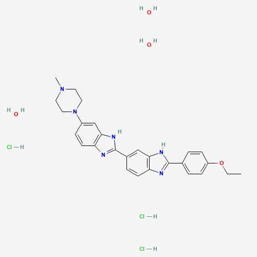
Hoechst 33342 staining solution (ready to use)
CAS number:881691-13-0 molecular formula:C27H28N6O•3HCl•3H2O
overview
compound introduction
Specs
| Chinese alias | - | ||
| English alias | BisBenzimide H33342 | HOE 33342 | ||
| CAS number | 881691-13-0 | molecular formula | C27H28N6O•3HCl•3H2O |
| molecular weight | 616.00 g/mol | Exact mass | 5ug/ml in H2O |
| PSA | - | logp | - |
numbering system
No numbering system information yet
physicochemical properties
No physical and chemical property information yet
Safety information
No safety information yet
Production methods and uses
No production method and use information yet
Related
upstream information
No upstream information yet
downstream information
No downstream information yet
MSDS
Found 0 shareMSDS